Method
The illustration is built from the medical files, then checked against the radiology.
Work begins with the medical reports: the emergency-room discharge, imaging reports, surgical reports, and consultations for future surgeries. From there we pull the imaging that is critical to the foundation of the case — images that directly correlate to the radiology reports or to the post-operative condition of the client.
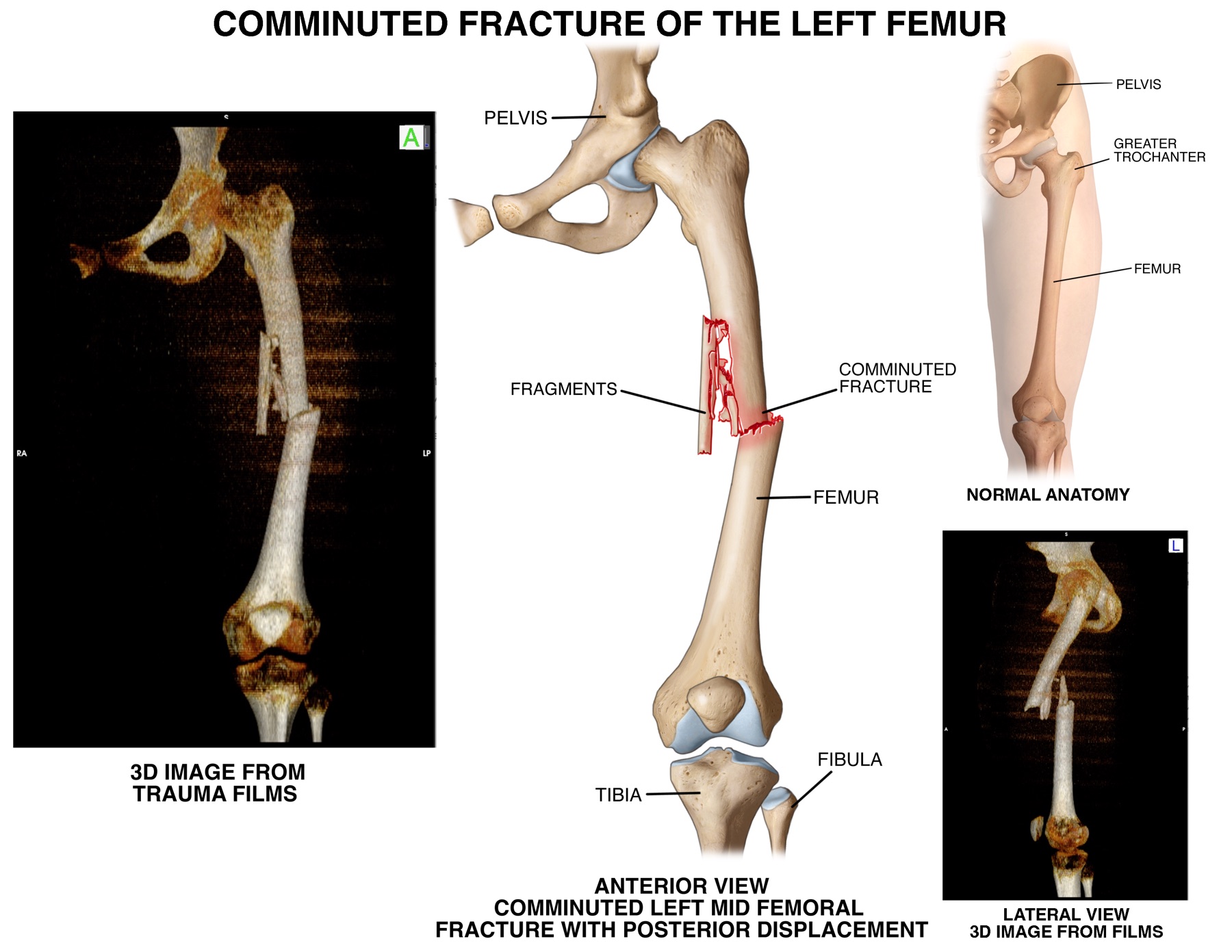
CT and MRI studies often contain hundreds of slices. We format those studies into 3D imaging that can give a 360° view of a fracture or a bird’s-eye view of a disc bulge. After the elements are gathered, we assemble the imaging, the reported facts, and photographs when they exist, and conceptualize a custom graphic.
Custom medical illustrations are built using NCG’s archive of thousands of illustrations from more than 35 years in the industry, together with current professional software. Fractures and soft-tissue injuries are traced accurately. The finished illustration is placed next to the radiology slide so an expert can see, at a glance, that the custom graphic correlates to the imaging.
The result is a medical illustration that is accurate, descriptive, and understandable to a layperson. Trial lawyers can feel confident with a custom exhibit created by NCG because every element of the case is brought together in one graphic that tells the story of the plaintiff’s injuries.